
Introduction: A Mystery Inside the Brain
The human brain weighs roughly three pounds, contains nearly one hundred billion neurons, and silently directs every heartbeat, spoken sentence, and cherished memory. It is also an organ that cannot feel pain directly, as brain tissue itself lacks sensory pain receptors. When an abnormal cellular growth emerges within this delicate environment, the initial clues rarely arrive with dramatic announcements. Instead, they often masquerade as everyday fatigue, subtle vision blurs, or minor slips of memory.
This creates one of modern medicine’s most profound investigative challenges: determining whether a subtle neurological shift is a harmless, transient issue or the first sign of an underlying tumour. Understanding brain cancer begins with dispelling fear through scientific clarity. Symptoms alone cannot confirm a diagnosis, and abnormal growths inside the skull vary tremendously in their biology, behavior, and treatability.
This comprehensive guide investigates the true nature of brain cancer. Below, you will discover how doctors differentiate benign growths from aggressive malignancies, which warning signs warrant medical attention, how modern diagnostic tools unravel the mystery, and what the latest evidence reveals about treatment options and daily living.
Table of Contents
What Is Brain Cancer?
At its most fundamental biological level, cancer occurs when cells escape normal growth controls, multiplying uncontrollably and forming a mass known as a tumour. When this uncontrolled replication occurs within the central nervous system, it presents unique anatomical challenges. Because the adult skull is an unyielding, rigid vault of bone, any expanding mass increases pressure inside the head, regardless of how fast or slow it grows.
Medical science divides brain tumours into two primary categories:
- Primary Brain Tumours: These growths originate directly within the tissues of the brain or its immediate surroundings, such as the meninges (the protective membranes encasing the brain) or cranial nerves. Primary tumours can be either non-cancerous (benign) or cancerous (malignant). According to the National Cancer Institute, primary brain tumours are classified according to the specific cells from which they arise, such as astrocytes, oligodendrocytes, or ependymal cells.
- Secondary or Metastatic Brain Tumours: These are tumours that develop when cancer cells break away from a primary site elsewhere in the body, such as the lungs, breasts, colon, or skin (melanoma), and travel through the bloodstream to seed new tumours in the brain. Metastatic tumours are far more common than primary brain tumours. Crucially, a lung cancer that spreads to the brain remains biologically lung cancer, and doctors treat it according to the biological profile of the primary tumour.
The distinction between benign and malignant tumours inside the skull differs from tumours elsewhere in the body. A benign brain tumour does not contain cancerous cells and does not invade surrounding healthy tissue or spread to distant organs. However, because space within the cranium is strictly limited, even a slow-growing, histologically benign tumour can compress vital structures, disrupt neurological function, and require urgent intervention. Malignant brain tumours, by contrast, grow rapidly, invade adjacent brain parenchyma aggressively, and carry a high likelihood of recurrence.
Tumour grade, anatomical location, and molecular markers dictate both the severity of the condition and the direction of clinical care.
Warning Signs That Deserve Attention
Because different areas of the brain govern distinct neurological tasks, the signs of a brain tumour depend largely on where the mass is located and how quickly it causes local irritation or general intracranial pressure.
Common warning signs include:
- Persistent or Progressive Headaches: Tumour-related headaches often worsen over time, feel distinctly different from past tension headaches, and may be most intense early in the morning or when coughing, bending, or straining.
- Seizures: A new seizure in an adult with no prior history of epilepsy is one of the most prominent presenting indicators of a brain tumour.
- Focal Weakness or Numbness: Gradual loss of sensation or strength on one side of the face, an arm, or a leg.
- Vision or Hearing Changes: Unexplained double vision, loss of peripheral vision, or sudden hearing difficulties.
- Speech and Language Difficulties: Stumbling over common words, difficulty comprehending normal conversation, or slurred articulation.
- Balance and Coordination Deficits: Unsteadiness on one’s feet, clumsy hand movements, or unexplained vertigo.
- Cognitive or Behavioral Shifts: Subtle changes in personality, apathy, unusual irritability, or uncharacteristic confusion.
Context is essential. Having a persistent headache does not mean you have a brain tumour. Tension headaches, migraines, dehydration, sinus infections, and stress account for the overwhelming majority of head pain.
As outlined by the NHS guidelines on brain tumours, you should seek a non-urgent medical evaluation if you experience persistent, unexplained, or gradually worsening symptoms over several weeks.
Seek immediate emergency medical attention if you experience:
- A first-time seizure or convulsion.
- Sudden onset of severe weakness, numbness, or paralysis on one side of the body.
- Sudden loss of speech, vision, or balance.
- An abrupt, explosive headache unlike anything previously experienced.
How Doctors Investigate the Mystery
Uncovering the cause of neurological symptoms resembles a methodical forensic investigation. Clinicians move systematically through several phases of objective testing before drawing conclusions.
Diagnostic Pathway:
[Comprehensive Clinical & Neurological Exam]
                    │
                    ▼
[High-Resolution Neuroimaging (MRI with Contrast / CT)]
                    │
                    ▼
[Neurosurgical Consultation & Pre-Operative Planning]
                    │
                    ▼
[Surgical Biopsy & Histopathological / Molecular Analysis]
1. Clinical and Neurological Examination
The evaluation begins with a comprehensive physical and neurological exam. The physician assesses cranial nerve function, pupil reactivity, visual fields, muscle strength, deep tendon reflexes, sensory perception, gait, and coordination. An ophthalmoscope examination of the back of the eye can detect swelling of the optic nerve (papilledema), a physical indicator of elevated intracranial pressure.
2. Neuroimaging
If the neurological examination reveals anomalies, advanced imaging is the next step:
- Magnetic Resonance Imaging (MRI): The gold standard for cranial diagnostics. Using powerful magnetic fields and radio waves, an MRI with intravenous gadolinium contrast generates cross-sectional images of brain soft tissue, identifying tumour dimensions, edema, and structural displacement.
- Computed Tomography (CT) Scan: CT imaging uses specialized X-rays. While less detailed for brain soft tissue than an MRI, CT scans are rapid, widely accessible in emergency rooms, and effective at detecting acute bleeding, calcifications, and structural bone erosion.
Diagnostic Limitation: Imaging alone cannot provide a definitive cancer diagnosis. A bright spot or mass on an MRI can represent a primary brain tumour, a metastasis, an infection, an abscess, a localized stroke, or an inflammatory condition such as multiple sclerosis.
3. Biopsy and Molecular Profiling
A definitive diagnosis requires examining physical tissue under a microscope. A neurosurgeon obtains a tissue sample through either a stereotactic needle biopsy (using computerized three-dimensional spatial coordinates) or an open surgical resection.
Pathologists analyze the cells to establish the tumour type and grade. Crucially, modern diagnostics also involve molecular and genetic profiling. Testing for mutations such as IDH1/IDH2 and checking the methylation status of the MGMT gene promoter provide vital clues regarding how the tumour developed and which therapies offer the highest likelihood of response.
Brain Cancer Treatment: What Are the Options?
No single treatment protocol applies to every patient. Care is tailored to the exact cellular type, grade, molecular profile, and anatomical location of the tumour, alongside the patient’s age and baseline physical health.
The American Cancer Society details several standard and emerging therapeutic approaches:
Surgery
Surgery is frequently the initial and most critical step. When a tumour is located away from vital functional regions, the neurosurgeon aims for maximal safe resection: removing as much tumour tissue as possible without causing permanent neurological damage. Modern operating rooms employ advanced tools such as intraoperative MRI, stereotactic neuronavigation, functional brain mapping, and awake craniotomies to protect areas governing speech and movement. If complete removal is impossible due to proximity to critical structures, a partial removal (debulking) can still reduce intracranial pressure and enhance the effectiveness of subsequent therapies.
Radiotherapy
Radiation therapy utilizes focused, high-energy beams (such as photons or protons) to damage the DNA of cancer cells, inhibiting their ability to divide. Approaches include:
- External Beam Radiation Therapy (EBRT): Directs radiation to the entire tumour bed and a surrounding safety margin over several weeks.
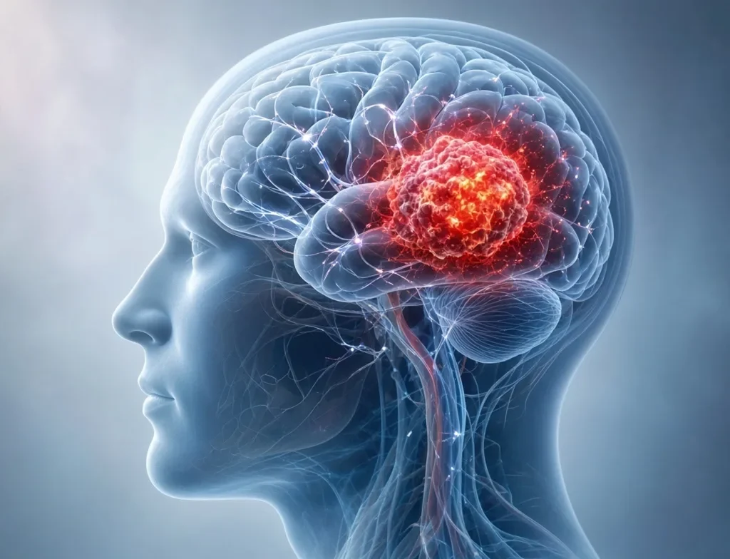
- Stereotactic Radiosurgery (SRS): Despite its name, this is not open surgery. Systems like Gamma Knife and CyberKnife focus hundreds of sub-millimeter radiation beams directly onto a precisely mapped lesion in a single session or a few sessions, sparing adjacent healthy tissue.
Chemotherapy
Chemotherapy uses cytotoxic medications to destroy rapidly dividing cells. A significant challenge in treating brain tumours is the blood-brain barrier, a natural protective network of tightly joined blood vessel cells that shields brain tissue from harmful toxins while blocking many conventional medications. Certain medications, such as temozolomide, cross this barrier effectively and are widely utilized in treating high-grade primary brain malignancies.
Targeted Therapies and Immunotherapy
Targeted therapies focus on specific molecular vulnerabilities within cancer cells, such as abnormal proteins that fuel growth, while minimizing injury to normal tissues. Immunotherapies, including immune checkpoint inhibitors, aim to help the body’s natural immune system recognize and attack malignant cells. While immunotherapy has transformed the management of several systemic cancers, researchers continue to refine its application against the immune-suppressive environment of brain tumours through active ClinicalTrials.gov studies.
Brain Cancer Facts Versus Common Myths
Medical misunderstandings generate unnecessary panic and lead to delayed care. Separating myth from established clinical evidence brings critical balance to health decisions.
| Common Myth | Evidence-Based Reality |
| Myth 1: Every brain tumour is cancerous. | Reality: Approximately two-thirds of all primary brain tumours diagnosed in adults are non-cancerous (benign), such as most meningiomas and pituitary adenomas. |
| Myth 2: Every persistent headache signals brain cancer. | Reality: Primary brain tumours are rare, whereas primary headache disorders (such as migraines and tension headaches) affect over a billion individuals worldwide. |
| Myth 3: All brain cancers behave identically. | Reality: Over 120 distinct central nervous system tumour subtypes exist, ranging from slow-growing low-grade lesions to aggressive, fast-moving glioblastomas. |
| Myth 4: One standard treatment cures all cases. | Reality: Treatment plans must be highly customized based on molecular markers, age, tumour location, and functional goals. |
| Myth 5: Online symptom checkers can diagnose a tumour. | Reality: Digital algorithms cannot examine retinas, check deep tendon reflexes, or interpret neuroimaging. Only trained clinicians can formulate a diagnosis. |
| Myth 6: Survival statistics dictate an individual’s future. | Reality: Published survival curves represent historical population averages from years past and cannot predict an individual patient’s personal outcome. |
A Practical Framework for Patients and Families
When neurological concerns arise, navigating the medical system can feel overwhelming. Patients and families can regain agency by applying the four-step Notice, Record, Consult, Verify framework.
       [ NOTICE ]
Unusual, persistent, or worsening functional changes
          │
          ▼
       [ RECORD ]
Document timing, triggers, patterns, and questions
          │
          ▼
       [ CONSULT ]
Seek formal medical evaluation; avoid self-diagnosis
          │
          ▼
       [ VERIFY ]
Confirm guidance via qualified multidisciplinary experts
- Notice: Pay calm, objective attention to changes that are progressive, persistent, or uncharacteristic. Isolated sensations that resolve quickly are rarely alarming; sustained patterns warrant attention.
- Record: Maintain an organized log. Document the precise time a symptom began, its duration, what made it better or worse, and how it affected daily functions. Note current medications, supplements, and family health history.
- Consult: Schedule an evaluation with a primary care physician or neurologist rather than relying on search engines. Present your clear symptom log to give your clinician an objective foundation for decision-making.
- Verify: If testing reveals a mass, verify all findings with qualified medical specialists, including neurosurgeons and neuro-oncologists. Seek second opinions at high-volume comprehensive cancer centers, confirm the rationale behind every recommended intervention, and inquire about active clinical trial opportunities.
Important Notice: This framework serves purely as an educational communication aid between patients and physicians. It is not an instrument for clinical self-diagnosis or medical triage.
What the Evidence Really Tells Us
Interpreting cancer statistics requires careful context. According to global surveillance data compiled by the International Agency for Research on Cancer (IARC), primary malignant brain and central nervous system tumours account for less than two percent of all cancer diagnoses worldwide. They remain relatively rare conditions compared to breast, prostate, lung, and colorectal cancers.
When patients review five-year survival figures, they are looking at retrospective population data. These figures describe individuals diagnosed and treated five to ten years ago under previous standards of care. They cannot capture recent advancements in surgical precision, molecularly guided radiotherapy, or modern systemic treatments.
Furthermore, broad statistical categories group diverse patients together. An individual’s personal trajectory depends on:
- Specific genetic mutations (IDH status, MGMT promoter status, 1p/19q co-deletion).
- Extent of surgical removal.
- Baseline functional score (performance status).
- Age and overall physical resilience.
Population statistics are administrative measures for public health researchers. They are never a personalized prognosis for an individual patient.
Real-World Application: From Fear to Informed Action
To understand how clear communication improves clinical care, consider this illustrative, hypothetical scenario:
Illustrative Example:
David, a 46-year-old high school teacher, noticed recurring morning nausea and subtle difficulty reading text on the left side of his computer monitor over a three-week period. He also experienced a mild, dull headache that tended to ease by midday.
Rather than cycling through alarming internet searches, David activated a simple symptom log. For seven days, he noted the time his headaches occurred, rated their intensity, and documented specific instances where his peripheral vision seemed impaired.
When David met with his primary care doctor, he presented his structured log rather than vague recollections. Recognizing a clear pattern of localized neurological symptoms, the physician performed an initial visual field examination and ordered a brain MRI with contrast.
Because David arrived prepared, his physician had the objective evidence required to justify rapid neuroimaging, cutting through weeks of uncertainty and ensuring an immediate referral to a neurosurgical team.
What Readers Should Do Next
If you or a loved one are coping with neurological symptoms or confronting a recent diagnosis, take these practical, constructive steps:
- Avoid self-diagnosis based on isolated symptoms: The central nervous system produces overlapping signals for hundreds of non-cancerous conditions.
- Schedule a clinical evaluation for persistent symptoms: Arrange an appointment with a primary care clinician if changes persist or worsen over days or weeks.
- Seek emergency services for red-flag symptoms: Go directly to an emergency department if there is an unexplained seizure, sudden limb paralysis, or sudden loss of speech or vision.
- Engage in shared medical decision-making: Write down questions before clinical visits, bring a trusted advocate to take notes, and ask specialists to explain the goals, benefits, and side effects of each proposed strategy.
- Rely on accredited medical institutions: Base your understanding on established cancer organizations and verified clinical guidelines rather than unverified online forums or unproven alternative remedies.
Conclusion: Knowledge Can Replace Confusion With Hope
The brain remains the most intricate organ known to science, and a potential diagnosis involving it is naturally intimidating. Yet medical understanding of the brain has advanced dramatically. Where clinicians once relied solely on broad observations, today’s teams deploy high-definition imaging, microsurgical mapping, and molecular profiling to understand and treat abnormal cellular growths with remarkable precision.
Facing the unknown can generate confusion, but sound scientific evidence provides a steady compass. By knowing which warning signs require medical attention, collaborating openly with qualified specialists, and making decisions based on verified clinical facts, patients and their families can navigate neurological health challenges with confidence, dignity, and genuine hope.
Frequently Asked Questions
What is the difference between a brain tumour and brain cancer?
A brain tumour refers to any abnormal mass of cells within the cranial cavity, including non-cancerous (benign) and cancerous (malignant) growths. Brain cancer specifically refers to malignant tumours that grow rapidly and invade surrounding healthy brain tissue.
What symptoms can occur with brain cancer?
Symptoms vary depending on the tumour’s location and size. Common signs include persistent or worsening morning headaches, new-onset seizures, unexplained nausea or vomiting, gradual weakness or numbness on one side of the body, balance difficulties, and changes in speech, vision, or personality.
Does a headache automatically mean brain cancer?
No. The vast majority of headaches stem from common, non-cancerous conditions such as tension headaches, migraines, eye strain, dehydration, or sinus pressure. Tumour-related headaches are relatively uncommon and typically accompany other progressive neurological deficits.
How do doctors diagnose brain cancer?
Diagnosis involves a thorough neurological examination, followed by high-resolution neuroimaging, most commonly an MRI with intravenous contrast. A definitive diagnosis requires a surgical biopsy to analyze tissue structure and genetic biomarkers under a microscope.
What treatments may be available for brain cancer?
Treatment plans are individualized and commonly include surgical resection to remove as much tumour as safely possible, radiation therapy to target residual cells, chemotherapy to inhibit cellular replication, and targeted or immunotherapy agents in selected cases.
Can brain cancer be cured?
Curability depends on tumour type, histological grade, molecular profile, and location. While some low-grade or localized primary tumours can be successfully eliminated or controlled for decades, aggressive high-grade tumours often recur. In such cases, modern medicine focuses on extending survival and preserving quality of life through comprehensive multidisciplinary care.
Where can patients and families find trustworthy information?
Reliable information is available from major, accredited cancer institutions, including the National Cancer Institute, the American Cancer Society, the NHS, and specialized organizations such as the American Brain Tumor Association and the National Brain Tumor Society.
Medical Information Disclaimer
This article is published exclusively for educational and informational purposes and does not constitute formal medical advice, diagnosis, or treatment recommendations. Always consult a qualified physician or healthcare provider regarding any medical condition, persistent symptoms, or treatment options. Never disregard professional medical advice or delay seeking care because of information read in this publication.