

It starts with something deceptively ordinary. A tiny, painless bump sits quietly on an arm or leg. It looks like an insect bite or a minor scratch from a garden shrub. There is no fever, no burning sensation, and no sudden pain to sound an alarm.
Days turn into weeks. Instead of fading, the spot widens beneath the surface. The tissue underneath begins to soften, yet the skin above feels almost numb.
How can a skin condition cause significant destruction beneath the surface without causing agony first?
This quiet progression defines one of medicine’s most fascinating microbial puzzles: Buruli ulcer. By understanding how this uncommon infection operates, we can strip away fear, recognise early warning signs, and protect our communities through prompt medical care.
Table of Contents
What Is Buruli Ulcer?
Buruli ulcer is a chronic, debilitating infection of the skin and soft tissues caused by an environmental bacterium named Mycobacterium ulcerans.
Biologically, this organism belongs to the same family of bacteria responsible for tuberculosis and leprosy. Unlike its airborne relatives, M. ulcerans targets the fatty tissue right beneath our skin.
The World Health Organization (WHO) classifies Buruli ulcer as a neglected tropical disease. While it is most frequently reported in rural communities across West and Central Africa, cases also occur in parts of South America, the Western Pacific, and coastal southeastern Australia.
No matter the geography, the infection thrives near slow-moving water bodies, wetlands, and temperate coastal regions.
Why This Disease Can Be Misleading
Most acute skin infections announce themselves with fierce inflammation. When common bacteria invade a cut, the immune system rushes in, causing heat, swelling, redness, and sharp throbbing pain.
Buruli ulcer breaks that fundamental rule.
The secret lies in a unique lipid toxin produced by Mycobacterium ulcerans called mycolactone. Mycolactone possesses two dangerous superpowers:
- It destroys tissue: It causes cell death (necrosis) in subcutaneous fat and collagen.
- It silences pain: It interacts with nerve signaling pathways and acts as a local anaesthetic while suppressing local immune responses.
Because the early lesion rarely hurts, many individuals dismiss it as harmless. Weeks or months pass before an open ulcer forms, allowing the bacteria to damage deeper layers unnoticed.
5 Signs Worth Knowing
Recognising early symptoms makes all the difference between a straightforward medical recovery and long-term joint complications. In our broader series on identifying infectious skin conditions, early observation remains the foundation of recovery.
Watch for these five progressive presentations:
- A firm, painless nodule: A small lump under the skin, often mistaken for a harmless cyst or bug bite.
- A plaque: A raised, broad, painless patch of skin that feels thicker and firmer than surrounding tissue.
- Localised oedema: A diffuse, painless swelling spreading across an entire limb or facial region.
- Undermined ulcer edges: As the fatty tissue underneath dies, the skin breaks open to reveal a crater with characteristic sunken, hanging edges.
- Absence of systemic fever: Patients generally feel entirely well systemically, which frequently delays an urgent clinic visit.
How Buruli Ulcer Spreads
Despite decades of investigation by international research teams, the exact transmission chain of Mycobacterium ulcerans remains partly mysterious.
We know that Buruli ulcer is not contagious from person to person. You cannot catch it by shaking hands with an affected individual or sharing a household meal.
Instead, transmission links directly to the environment:
- Insect vectors: In parts of southeastern Australia, verified studies have detected the bacteria in native possums and mosquitoes, demonstrating that mosquito bites can transmit the pathogen to humans.
- Environmental abrasions: In rural tropical zones, tiny cuts, scratches, or punctures acquired while farming, fishing, or washing in swampy waters can allow bacteria from damp soil or biofilm to enter open skin.
What Happens Inside the Skin?
To understand how the bacteria work, picture an iceberg. The visible mark on the skin is just the tip.
Once Mycobacterium ulcerans settles inside the warm, lipid-rich subcutaneous fat, it starts producing mycolactone. The toxin spreads outward like an invisible ring, dissolving fat cells and deadening the tiny nerve endings that relay distress signals to the brain.
[Bacterial Entry via Skin Break or Vector]
                 │
                 ▼
[Invasion of Subcutaneous Fatty Tissue]
                 │
                 ▼
[Production of Mycolactone Toxin]
   ├── Disables local immune cells
   ├── Shuts down local pain signals (Numbness)
   └── Dissolves subcutaneous fat layer
                 │
                 ▼
[Overlying Skin Collapses → Classic Ulcer Formation]
Without early intervention, the collapse of underlying fat eventually starves the top layers of skin of blood supply, causing the surface to slough away into an open, expanding ulcer.
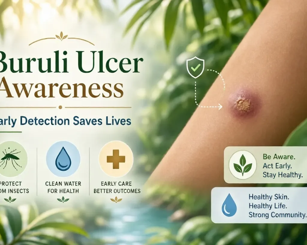
A Short Medical Mystery
The following scenario is a fictional, illustrative case study designed to show how Buruli ulcer typically develops and why early clinical evaluation is essential.
A Small Mark That Would Not Explain Itself
Thomas, a 42-year-old landscaper living along a coastal river basin, noticed a pale, nickel-sized lump on his left forearm. He assumed it was a harmless spider bite from clearing brush near stagnant drainage pools.
Three weeks passed. The bump did not itch or sting, but it steadily widened into a firm, raised plaque. Thomas continued working, reassured by the lack of pain. “If it were dangerous, it would hurt,” he told his partner.
By week six, the center of the firm plaque softened and gave way, exposing a shallow ulcer with rolled, undermined margins. Surprised by the lack of pain despite the visible sore, Thomas scheduled an appointment with a dermatologist.
Suspecting an atypical mycobacterial infection, the doctor performed a swab and a fine-needle biopsy for Polymerase Chain Reaction (PCR) testing.
Within days, the laboratory confirmed the presence of Mycobacterium ulcerans.
Because Thomas presented before deep tissue, muscle, or tendon involvement occurred, his clinical team initiated a verified eight-week oral antibiotic regimen combining rifampicin and clarithromycin, supplemented with sterile dressings. Within two months, the infection cleared, leaving a clean scar and full range of motion in his arm.
How Doctors Diagnose It
Diagnosing Buruli ulcer demands clinical suspicion combined with laboratory confirmation. Because the lesions resemble fungal infections, leishmaniasis, or diabetic foot sores, healthcare workers rely on four primary methods endorsed by global health bodies:
- PCR (Polymerase Chain Reaction): The gold standard. PCR tests target the specific genetic sequence IS2404, providing definitive confirmation within forty-eight hours.
- Direct Smear Microscopy: A fast, low-cost clinic test that stains skin swabs with Ziehl-Neelsen dye to identify acid-fast bacilli under a microscope.
- Histopathology: Microscopic examination of a small skin biopsy to check for fat necrosis and clusters of extracellular bacteria.
- Bacterial Culture: Growing the bacteria in an incubator. Because M. ulcerans multiplies exceptionally slowly, cultures can take eight to twelve weeks to yield results, making it less practical for immediate treatment decisions.
What Modern Treatment Can Do
There is excellent news: Buruli ulcer is completely curable.
Years ago, treatment often required extensive surgery to excise large areas of infected tissue. Today, health guidelines from the World Health Organization (WHO) prioritise oral antimicrobial therapies.
       EARLY MEDICAL TREATMENT
                  │
  ┌───────────────┴───────────────┐
  ▼                               ▼
[Oral Antibiotics]        [Supportive Care]
• Rifampicin              • Sterile wound dressings
• Clarithromycin          • Physiotherapy (joint mobility)
• Daily for 8 weeks       • Nutritional support
- Standard Medication: Most patients complete an eight-week daily course of oral antibiotics, typically combining rifampicin with clarithromycin. This non-invasive combination cures over 90% of early-stage infections.
- Wound Management: Clean, moist wound dressings protect the skin from secondary bacterial invaders, promoting tissue regeneration.
- Physiotherapy: When an ulcer forms across an elbow, knee, or wrist, regular mobility exercises prevent permanent scarring and joint contractures.
- Surgical Debridement: Reserved primarily for advanced cases to clear away extensive necrotic tissue or apply skin grafts to accelerate healing.
For readers seeking further context on immune responses during antibiotic therapy, explore our guide on understanding antibiotic treatments and tissue recovery.
How Prevention Can Help
Because we do not yet possess a dedicated Buruli ulcer vaccine, prevention relies on practical barriers:
- Wear protective clothing: Long pants and long-sleeved shirts reduce skin abrasions while gardening, farming, or exploring coastal wetlands.
- Apply insect repellent: In regions where mosquito transmission is confirmed, using DEET- or picaridin-based repellents lowers bite risks.
- Wash cuts immediately: Clean any scrape or insect bite with clean water and soap, and cover it with a protective bandage until closed.
- Avoid washing directly in stagnant waters: Rural communities should utilise boreholes and protected water sources whenever available.
When Should You Seek Medical Care?
You should consult a healthcare professional whenever you notice:
- Any skin nodule, firm patch, or swelling that persists for more than two weeks without resolving.
- An open sore that shows no sign of healing, especially if it does not hurt as much as its appearance suggests.
- A wound that develops sunken, ragged, or undermined edges.
- Persistent skin changes after traveling to wetlands, endemic rural areas, or coastal regions.
Prompt evaluation prevents extensive tissue loss and preserves normal physical mobility.
Frequently Asked Questions
What is Buruli Ulcer?
Buruli ulcer is a chronic, progressive infection of the skin and underlying fat tissue caused by an environmental bacterium called Mycobacterium ulcerans. It causes painless lumps, plaques, and eventual ulcers.
Is Buruli Ulcer contagious?
No. Buruli ulcer is not transmitted directly from person to person. Transmission occurs through exposure to contaminated environmental sources, damp soil, or bites from insect vectors like mosquitoes in endemic locations.
What causes Buruli Ulcer?
It is caused by Mycobacterium ulcerans, an organism that secretes a destructive toxin called mycolactone. This toxin dissolves subcutaneous fat cells while dulling local nerve responses.
What are the early signs of Buruli Ulcer?
Early signs typically include a firm, painless swelling (nodule), a raised hardened area (plaque), or broad painless swelling (oedema), usually situated on the arms or legs.
Can Buruli Ulcer be treated?
Yes. An eight-week course of oral antibiotics (such as rifampicin and clarithromycin), paired with proper wound dressing and physical therapy, cures the vast majority of cases without major surgery.
How is Buruli Ulcer diagnosed?
Clinicians examine the wound and confirm the diagnosis through laboratory testing. Polymerase Chain Reaction (PCR) testing is the most accurate, rapid diagnostic tool used globally.
Can Buruli Ulcer be prevented?
While there is no specific vaccine, you can reduce risk by wearing protective clothing near wetlands, applying insect repellent, and cleaning and covering skin scrapes promptly.
When should someone see a doctor about a suspicious skin lesion?
Seek a medical evaluation if you have an unusual bump, swelling, or open sore that fails to heal after two to three weeks, particularly if it feels unusually painless or follows outdoor activity in wetland areas.
Medical Disclaimer: This article is intended strictly for educational and informational purposes. It does not constitute medical advice, diagnosis, or treatment. Always consult a qualified physician or healthcare provider regarding any suspicious skin lesion or persistent medical condition.