
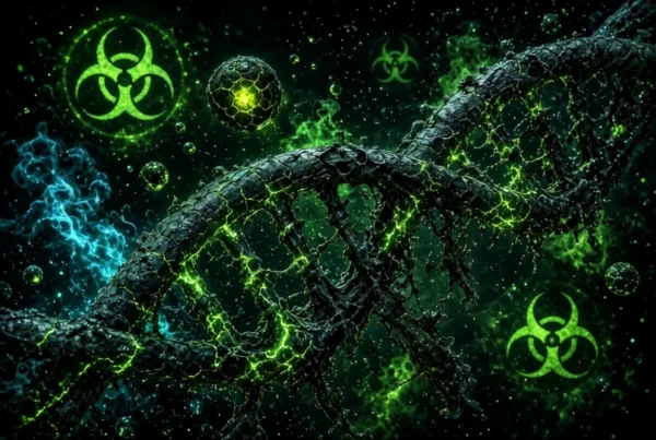
Toxic DNA :Inside a single living cell, everything looks calm until it suddenly is not. Metabolic engines hum smoothly, proteins fold along delicate chemical tracks, and the double helix sits protected inside the nucleus. Then an unseen threat slips across the outer membrane. A cascade of unstable molecules floods the interior, stripping electrons from vital structures and tearing through the cell scaffold. Deep within the core, the genetic blueprint fractures.
Warning signals fire across molecular pathways. Enzymes rush to stabilize the breaks, working against a ticking biological clock. If the repairs hold, the cell resumes its normal duties. If the destruction is too vast, the cell must execute a dramatic choice: dismantle itself quietly or rupture, triggering widespread inflammation across neighboring tissue.
This silent struggle happens millions of times every day. It represents the thin line between survival and systemic breakdown, defined by two critical concepts in cell biology: cytotoxicity and Toxic DNA damage.
Table of Contents
What Is Cytotoxicity?
Cytotoxicity describes the quality of being toxic to cells. When a chemical substance, environmental agent, or immune cell causes cellular injury, it exerts a cytotoxic effect. This is not always a slow, passive decay. Depending on the concentration and nature of the agent, toxicity can disrupt nutrient transport, rupture outer membranes, or paralyze basic energy production.
In medical research, measuring cytotoxicity is a fundamental baseline. It helps toxicologists determine whether an experimental drug destroys healthy human tissue, or conversely, whether an oncology therapy effectively kills malignant tumors.
Why Cells Become Vulnerable
Cells are remarkably resilient, yet their internal balance is delicate. Cellular injury begins when external stressors overwhelm baseline regulatory defenses.
Several factors make cells vulnerable to toxic events:
- Membrane destabilization: Detergents, solvents, or lipid-destroying toxins strip the outer phospholipid bilayer, causing internal components to leak out.
- Metabolic disruption: Heavy metals and biological poisons can disable critical enzymes, freezing energy production in seconds.
- Oxidative overload: Excessive reactive oxygen species overpower native antioxidant networks, reacting indiscriminately with structural lipids and proteins.
- Impaired waste clearance: When damaged proteins accumulate faster than lysosomes and proteasomes can recycle them, internal transport grids collapse.
When these vulnerabilities are exploited simultaneously, the structural integrity of the cell deteriorates rapidly.
How Toxic DNA Damage Begins
At the molecular level, severe cellular stress rarely stays confined to the cytoplasm. Highly reactive molecules, particularly reactive oxygen species (ROS) generated during toxic metabolic stress, migrate into the nucleus. Here, chemical attacks strike the genetic code directly.
When these molecules collide with the double helix, they alter nucleotide bases and sever the sugar-phosphate backbone. This chemical violence creates structural breaks across single strands or, more dangerously, breaks across both complementary strands. If an agent causes chemical cross-links between DNA and surrounding proteins, the replication machinery stalls entirely.
This acute genetic injury stalls the transcription of essential proteins. The cell suddenly finds its primary operating manual unreadable at the exact moment it needs emergency repairs.
The Cellular Repair Battle
Cells do not surrender to genetic injury without a fight. Evolution has equipped our biology with an intricate system called the Toxic DNA Damage Response (DDR).
Sensor proteins constantly cruise along chromatin fibers. When they spot a lesion, they halt the cell cycle at dedicated checkpoints. This pause prevents the cell from duplicating corrupted code.
Specialized molecular repair crews then deploy:
- Base Excision Repair (BER): Removes small, non-helix-distorting base lesions caused by oxidation or alkylation.
- Nucleotide Excision Repair (NER): Carves out bulky lesions, such as those caused by ultraviolet radiation.
- Double-Strand Break Repair: Employs either homologous recombination for error-free restoration or non-homologous end joining to fuse severed ends back together.
If the structural crisis is contained, checkpoint brakes release, and normal biological function returns.
When Toxic DNA Repair Fails
When cellular toxicity is too severe or sustained, repair mechanisms become overwhelmed. At this critical junction, the cell arrives at an evolutionary fork in the road.
If sensor proteins detect irreparable genetic destruction, they activate apoptosis, also known as programmed cell death. The cell systematically shrinks, condenses its chromatin, and packages its contents into neat apoptotic bodies that circulating immune cells ingest without disturbing surrounding tissue.
However, if toxicity causes immediate, catastrophic energy loss or direct membrane destruction, the cell undergoes necrosis. The cell swells, its outer membrane bursts, and intracellular toxins spill into surrounding tissue, provoking acute inflammation.
In worst-case scenarios, a damaged cell survives with flawed code. It bypasses cycle checkpoints, accumulating mutations and driving genomic instability, an established driver of chronic disease pathways.
The Mitochondrial Connection
Mitochondria are often described as cellular powerhouses, but during cytotoxic stress, they act as primary arbiters of life and death.
Under toxic conditions, calcium rushes into the mitochondrial matrix while reactive oxygen species accumulate along the inner membrane. This triggers the opening of the mitochondrial permeability transition pore (mPTP). When this pore opens, the electrical gradient required to generate adenosine triphosphate (ATP) collapses instantly.
Without ATP, cellular pumps stop working. Even more critically, the permeabilized mitochondria release cytochrome c into the cytosol. This single molecular event binds with local adapter proteins to assemble the apoptosome, activating executioner caspase enzymes that irrevocably commit the cell to destruction.
A Real Scientific Thriller: The Benzene Investigation
In the mid-twentieth century, industrial workplaces increasingly relied on benzene, a sweet-smelling solvent used widely in rubber fabrication, chemical synthesis, and petroleum refining. For years, workers handled benzene with minimal respiratory or skin protection, unaware of its invisible biological cost.
In the 1970s and 1980s, epidemiological surveys led by researchers at the National Cancer Institute and global health bodies began documenting abnormal rates of bone marrow failure and acute myeloid leukemia among exposed factory workers.
Biochemical investigations soon cracked the mystery: benzene itself was not the primary culprit. Once inhaled, the liver metabolizes benzene into phenolic metabolites, which travel directly to the bone marrow.
Benzene Inhalation ➔ Liver Metabolism ➔ Phenolic Metabolites ➔ Bone Marrow Toxicity ➔ Severe DNA Damage
Inside marrow stem cells, myeloperoxidase enzymes convert these metabolites into aggressive benzoquinones. These compounds generate immense oxidative stress, inhibit topoisomerase II, and induce severe, recurrent DNA damage in hematopoietic progenitors.
The cell repair systems were overwhelmed by the chronic toxicity, causing chromosome aberrations and catastrophic marrow suppression.
The scientific verification of benzene-induced cytotoxicity and genetic damage spurred landmark occupational safety regulations globally. Permissible exposure limits were slashed worldwide, establishing modern standards for biological monitoring and chemical risk assessment that protect millions of workers today.
How Scientists Detect Cytotoxicity
Modern laboratory testing relies on a suite of sensitive analytical assays to detect whether compounds harm cells:
| Assay Type | Target Biomarker | Scientific Purpose |
| MTT / WST-1 Assay | Mitochondrial Dehydrogenase | Measures overall metabolic activity and viable cell count |
| LDH Release Assay | Lactate Dehydrogenase | Detects physical rupture of the outer plasma membrane |
| DCFH-DA Staining | Intracellular Fluorophores | Quantifies reactive oxygen species and oxidative stress levels |
| Comet Assay | Electrophoretic DNA Migration | Visualizes single and double-strand genetic breaks directly |
| Annexin V / PI | Phosphatidylserine Externalization | Distinguishes early programmed apoptosis from late necrosis |
Using these multi-endpoint testing strategies, researchers assess pharmaceutical safety and characterize environmental toxins long before clinical trials begin.
Cytotoxicity Versus Genotoxicity
While these two terms frequently appear together in toxicology reports, they describe distinctly different biological events.
Cytotoxicity refers broadly to cellular destruction or impairment. A cytotoxic substance damages membranes, blocks metabolic pathways, or alters cellular architecture, potentially killing the cell outright.
Genotoxicity, on the other hand, specifically describes agents that damage genetic material, altering DNA sequence, structure, or chromosomal alignment.
A substance can be genotoxic without immediately killing the cell, allowing viable mutations to pass into subsequent generations. Conversely, an agent can be acutely cytotoxic (such as a strong acid or detergent) without directly binding to or chemically altering the DNA structure. Understanding this difference enables researchers to evaluate whether an exposure risks acute tissue damage or long-term mutagenic potential.
Why Everyday Exposure Matters
In daily life, human bodies encounter low concentrations of potential toxins continuously:
- Airborne pollutants: Fine particulate matter ($PM_{2.5}$) and volatile organic compounds carry oxidative chemicals into lung epithelial linings.
- Ultraviolet radiation: Solar radiation directly breaks DNA base pairings in skin cells while driving localized free radical generation.
- Dietary and lifestyle elements: Highly processed foods cooked at extreme temperatures produce advanced glycation end products, while alcohol metabolism yields cytotoxic acetaldehyde.
- Occupational materials: Industrial paints, chemical cleaners, and synthetic resins require vigilant handling to prevent chronic inhalation exposure.
Our tissues constantly process, neutralize, and clear these agents. Low-level exposures rarely cause immediate disease because evolutionary biological buffers actively neutralize threats before they reach critical toxicity thresholds.
How Cells Fight Back For Toxic DNA
The human body is not a passive target. Our tissues continuously deploy sophisticated protective networks to preserve biological homeostasis:
- Endogenous Antioxidants: Enzymes such as superoxide dismutase, catalase, and glutathione peroxidase neutralize reactive oxygen species before they strike vital organelles.
- Phase II Conjugation: Liver enzymes attach polar groups to toxic molecules, making them water-soluble for rapid excretion via bile or urine.
- Cell Cycle Arrest: Protein complexes pause cellular division the moment an anomaly is detected, ensuring flawed code is never copied.
- Immune Clearance: Specialized phagocytes continuously patrol tissues, engulfing cells that undergo apoptosis before they can rupture and cause inflammation.
These synchronized defenses ensure that occasional toxic challenges are resolved cleanly and quietly.
What Research Is Teaching Us
Current research in toxicology and cell biology is moving away from simple high-dose animal testing toward advanced human organ-on-a-chip models and computational systems biology.
Scientists now recognize that low-dose, chronic cellular toxicity operates along subtle molecular pathways rather than causing outright cell death. Modern studies focus on how minor, persistent inflammation, epigenetic changes, and repeated micro-injuries compromise tissue elasticity over decades.
By mapping the precise molecular transitions from cellular adaptation to cell death, researchers are discovering new therapeutic targets to protect heart tissue during chemotherapy and shield neurons from chronic degenerative decline.
Practical Takeaways Of Toxic DNA Damage
- Support natural detoxification: Drink adequate water, prioritize deep sleep, and consume dietary fiber to support healthy kidney, liver, and bowel clearance mechanisms.
- Emphasize dietary antioxidants: Whole fruits, vegetables, and leafy greens provide dietary polyphenols that bolster baseline cellular antioxidant defenses.
- Limit avoidable chemical exposure: Use proper personal protective equipment when handling industrial cleaners, chemical strippers, solvents, or garden pesticides.
- Protect your skin barrier: Use broad-spectrum sunscreen consistently to limit ultraviolet-induced radical formation in dermal layers.
- Avoid unnecessary panic: The human body manages thousands of minor metabolic and environmental stressors every hour through deeply robust, evolutionary repair mechanisms.
Frequently Asked Questions
What is cytotoxicity?
Cytotoxicity is the measure of how harmful or destructive a substance is to living cells. A cytotoxic agent can disrupt cell division, compromise outer membranes, or cause cell death.
What causes cytotoxicity?
It can be triggered by external chemicals, heavy metals, radiation, microbial toxins, synthetic drugs, or endogenous factors such as excessive free radicals and localized autoimmune responses.
Can cytotoxicity cause DNA damage?
Yes. Cytotoxic processes frequently create overwhelming oxidative stress and metabolic chaos, which damages chromosomes, fractures the double helix, and overwhelms natural cellular repair systems.
What is the difference between cytotoxicity and genotoxicity?
Cytotoxicity refers to general cell damage, functional impairment, or cell death. Genotoxicity refers specifically to chemical or physical damage directed at genetic material, regardless of whether the cell dies.
How is cytotoxicity measured in a laboratory?
Scientists evaluate cell viability using specialized assays, including MTT tests for metabolic energy, LDH leakage tests for membrane destruction, and the Comet assay to visualize genetic breaks.
Can damaged DNA be repaired?
Yes. Cells possess dedicated enzymatic systems, including base excision repair, nucleotide excision repair, and homologous recombination, designed to correct genetic lesions before cells divide.
When does cellular damage become dangerous?
Cellular injury becomes dangerous when exposure is so severe, prolonged, or frequent that internal repair machinery fails, resulting in extensive tissue necrosis, chronic inflammation, or permanent mutations.
Life at the microscopic scale is a balance between environmental wear and molecular resilience. Every second, our cells navigate toxic challenges, repair structural breaks, and protect our genetic heritage. By understanding the science behind cytotoxicity and cellular repair, we can better appreciate the invisible biological systems that sustain human health every day.
Faydalı. Muse Voice Transcribe: ile speech-to-text deneyin. Muse Voice Transcribe